
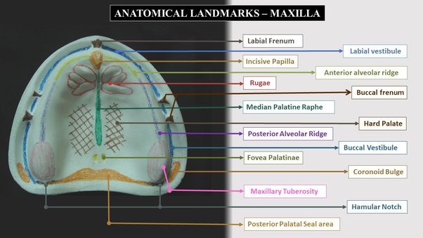
Anatomical Landmarks - Maxilla
Introduction
Anatomical landmarks are very important for fabricating dentures. Knowledge about anatomical landmarks helps to provide dentures with proper stability, retention and support.
Maxillary anatomical landmarks
Average available area of edentulous maxilla for denture is 24 sq. cm.
Supporting structures:
Hard palate
Residual alveolar ridge
Maxillary tuberosity
Labial frenum (1)
Labial vestibule
Buccal frenum (2)
Buccal vestibule
Hamular notch
Fovea palatine
Posterior palatal seal area
Stress bearing areas:
Primary: horizontal slopes of hard palate lateral to median suture
Secondary: residual alveolar ridge, rugae (anterior hard palate), maxillary tuberosity
Tertiary: posterolateral part of hard palate
Retentive areas:
Primary: Posterior palatal seal area
Secondary: posterolateral part of hard palate
Relief areas:
Incisive papilla
Mid palatine raphae
Fovea palatine
Cuspid eminence
Supporting structures
Hard palate:
Mucosa is keratinised.
Anterolaterally, it has rugae which is a stress bearing area.
Posterolaterally, submucosa has glandular tissue. It is a least resorptive area giving primary support.
Residual alveolar ridge:
It is a portion of alveolar process and it's soft tissue covering remains after extraction.
It resorbs with time and hence it is a secondary stress bearing area.
Maxillary tuberosity:
It is a bulbous extension of residual alveolar ridge in the 2nd molar and 3rd molar region. It rarely resorbs.
Rugae:
It is a raised area of dense connective tissue on the anterior third of the palate at an angle to the residual alveolar ridge.
It resists anterior displacement of denture.
Limiting structures
Labial frenum:
It is a fold of mucous membrane. It has no muscular attachment or no action. It inserts in a vertical direction.
Labial vestibule:
It is divided left and right by labial frenum.
It is a portion bound by residual alveolar ridge on one side and lips on outer side.
Orbicularis oris forms the outer surface of labial vestibule.
Buccal frenum:
It divides the vestibule into labial and buccal vestibules.
It may be one or two
It contains Caninus (levator anguli oris).
Buccinator pulls the frenum backward. Orbicularis oris pulls the frenum forward.
Buccal vestibule:
It extends from buccal frenum to hamular notch.
It is bounded by residual alveolar ridge medially and cheeks laterally.
The size of buccal vestibule is modified by:
1. Contraction of buccinator
2. Amount of bone loss in maxilla
3. Position of mandible
4. During mouth opening, coronoid process of mandible and the masseter muscle modify the size of buccal vestibule.
Hamular notch:
It is the distal limit of the buccal vestibule. It is present between maxillary tuberosity and pterygoid hamulus.
It aids in Posterior palatal seal.
Fovea palatine:
2 openings of palatal minor salivary glands present on either side of midline posterior ri junction of hard and soft palate.
It determines the posterior extent of upper denture. Denture should end 1-2 mm beyond it.
Posterior palatal seal area:
It consists of pterygomaxillary seal and post palatal seal.
Pterygomaxillary seal is formed from hamular notch and distobuccal surface of buccal vestibule.
Post palatal seal is the area between anterior and posterior vibrating lines in the soft palate.
Anterior vibrating line is an imaginary line formed between an immovable and slightly movable soft palate. It is usually cupid bow shaped. It is recorded by the patient saying ‘ah’ in short vigorous bursts or by Valsalva maneuver.
Posterior vibrating line is an imaginary line formed between a slightly movable and freely movable soft palate. It is present at the junction of aponeurosis of tensor veli palatini and soft palate muscles. It is a slightly curved line. It is located by instructing the patient to say ‘ah’ in a normal unexaggerated manner.
Incisive papilla:
It is a pear shaped elevation present in the anterior part of the edentulous maxilla. It lies just posterior to the maxillary central incisors. After extraction, it might migrate towards the residual alveolar ridge as the ridge resorbs. Beneath the incisive papilla, runs the nasopalatine vessels and nerves. It should always be relieved.
Mid palatine raphae:
It is the mucoperiosteum covering the median palatine suture. When it is prominent, it should be relieved.
2 comments
I'll post a few public awareness articles regularly!