
Anatomical Landmarks - Mandible
Introduction
Anatomical landmarks are very important for fabricating dentures. Knowledge about anatomical landmarks helps to provide dentures with proper stability, retention and support.
Mandibular Anatomical Landmarks
Limiting Structures:
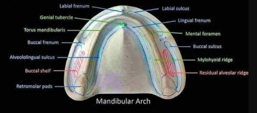
Labial frenum
Labial vestibule
Buccal frenum
Buccal vestibule - Masseteric notch
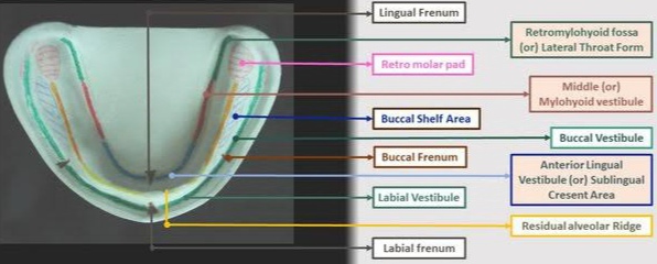
Retromolar pad
Lingual frenum
Alveololingual sulcus - retromylohyoid space
Mylohyoid ridge
Supporting Structures:
Buccal shelf area
Residual alveolar ridge
Stress bearing areas:
Buccal shelf area (primary)
Slopes of residual alveolar ridge (secondary)
Relief areas:
Crest of residual alveolar ridge
Genial tubercles
Mental foramen
Torus mandibularis (if present)
Limiting Structures
Labial Frenum:
It is an active band extending from the labial aspect of the ridge to the lip. It contains Orbicularis oris muscle.
Labial Vestibule:
It extends from labial frenum to buccal frenum. It is bounded by ridge on one side and lips on the other. The length and thickness of the labial flange of the denture occupying this space is important in influencing lip support and retention.
Buccal Frenum:
The fibers of the buccinator are attached to the frenum. It should be relieved to prevent displacement of the denture during function.
Buccal Vestibule:
It extends posteriorly from the buccal frenum till the retromolar region. It is bound by the residual alveolar ridge on one side and buccinator on the other side.
Masseteric Notch:
It is the influence of the masseter on the buccinator. If it is overextended in the mandibular denture, denture dislodges due to the action of masseter.
Retromolar pad:
It is a soft elevation of mucosa lying distal to the third molar. It has loose connective tissue with an aggregate of mucous glands. It is bounded medially by pterygomandibular raphae and superior constrictor, laterally by buccinator and posteriorly by tendon of temporalis. It plays a major role in peripheral seal and support.
Lingual Frenum:
It is a fold of mucous membrane extending along the floor of the mouth to the ventral surface of the tongue. It is activated on tongue movement. Relief should be provided in the anterior portion of the lingual flange. This anterior portion of the lingual flange is called sublingual crescent area.
Alveololingual Sulcus:
It extends from the lingual frenum to the retromylohyoid curtain. It is divided into 3 regions - anterior, middle, posterior
Anterior region:
It extends from lingual frenum to premylohyoid fossa where mylohyoid curves below the sulcus. The flange will be shorter anteriorly.
Middle region:
It extends from premylohyoid fossa to distal end of mylohyoid ridge. Due to the prominence of mylohyoid ridge and action of mylohyoid muscle, it is more shallow than other parts of sulcus.
Posterior region/ lateral throat form:
It extends from the end of mylohyoid ridge to retromylohyoid curtain. It is bounded posterolaterally by superior constrictor, posteromedially by palatoglossus and medially by tongue. It forms the S shaped curve of lingual flange of the mandibular denture.
Retromylohyoid area:
The muscles involved are superior constrictor and medial pterygoid.
Mylohyoid Ridge:
It is an irregular bony crest on the lingual surface of the mandible.
Supporting Structures
The support for a mandibular denture comes from the body of the mandible. The available denture-bearing area for an edentulous mandible is 12.25 sq. cm
Stress Bearing Area
Buccal Shelf Area:
It is the area between the buccal frenum and anterior border of Masseter. It is bounded medially by the crest of the ridge, distally by the retromolar pad, and laterally by the external oblique ridge.
It is a primary stress bearing area because it is present perpendicular to the occlusal forces.
Residual Alveolar Ridge:
It is made of spongy bone. The crest if the residual ridge should be relieved as stress in on it can cause resorption. The labial/buccal and lingual slopes of the ridge act as the secondary stress bearing area.
Relief Areas
Mental Foramen:
It is located in the lateral body of the mandible between the first and second premolar area. Mental nerve and blood vessels passes through it, hence it must be relieved.
Genial Tubercles:
These are pairs of 2 bong tubercles found anteriorly on the lingual surface of the mandible. The upper pair provide origin for genioglossus muscle, and the lower pair provide origin for geniohyoid muscle. It should be relieved.
Torus Mandibularis:
It is an abnormal bony prominence found on the lingual surface of the mandible near the premolar region bilaterally. It is covered by thin mucosa. Either it should be removed surgically or relieved during denture preparation according to its position and extent.
Post a Comment